A 30-year-old male construction worker presents to the clinic with a slight limp and discomfort in his right knee.
Sample Case
A 30-year-old male construction worker presents to the clinic with a slight limp and discomfort in his right knee. He tells the doctor that the injury has been recurring for the past three years, but was aggravated recently after a strenuous workout at his fitness club. He informs the doctor that he was performing squat exercises when he felt pain in his right knee. He continued to exercise for the remaining hour, but felt that the discomfort progressively increased as the exercise regime continued. He especially noticed it when climbing the stairs to leave the fitness centre. The patient now describes a grinding sensation in his right knee when he walks or runs. Physical examination reveals a subluxation at the right ilium, and subluxation of the right patella present through both static and motion palpation. Right quadriceps hypertonicity was also present. Neurological and radiological examinations were unremarkable.
Have you encountered a case like this? Are you comfortable assessing and correcting subluxations in the knee? In this edition of Technique Toolbox, we will look at the knee, and assess and correct for a superior lateral patella subluxation.
Before we begin, the doctor must initially ensure that all primary, secondary and tertiary subluxations of the spine have been assessed and corrected. In this particular case, an ilium subluxation was present on the right side. Therefore, the doctor must correct the subluxated ilium before moving on to the patellar problem.
To accurately detect and correct a subluxation of the patella, the doctor will perform the following regime.
 |
|
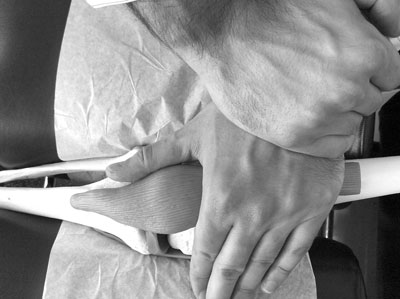
| Photo 1: Contacts for a superior lateral patella subluxation are displayed on a skeletal model. |
Analysis
- Patient presents with knee pain that increases in intensity during stair or hill climbing.
- Runners that perform a great deal of training on uneven terrain are especially susceptible to this type of subluxation.
Palpation
- The doctor can easily reproduce the pain through palpation of the lateral aspect of the patella and quadriceps tendon.
- The pain can also be reproduced through quadriceps resistance testing during the physical
- examination.
- Muscle resistance within the quadriceps also reveals a weakness in the vastus medialis.
Radiology
- If the injury is caused by impact or trauma, the doctor must take an X-ray to rule out the possibility of fracture or dislocation.
- As in all cases, a fracture or instability must be considered a contraindication to adjustment and must be referred out. Fortunately, this is a rare occurrence as most cases can be successfully corrected with chiropractic care.
- For clinical interest, the doctor should be aware that this subluxation pattern is often associated with Osgoode Schlatters, and will be detected on X-ray analysis.
- If X-ray analysis indicates no sign of fracture or instability, the doctor can continue to correct this subluxation pattern.
The above clinical findings indicate that the patella has subluxated superior and laterally.
The doctor must note that this condition is usually associated with findings in the spine, namely, an original posterior inferior (PI) ilium. It is not in the scope of this article to describe this particular subluxation in great detail. However, the doctor must be mindful that the PI ilium causes an excessive lengthening and subsequent neurological contraction on the quadriceps muscles, especially the rectus femoris. When there is a weakness in the vastus medialis, this excessive pull subluxates the patella superior and lateral. Thus, as stated earlier, the ilium subluxation must be corrected prior to correcting the patellar subluxation.
Correction
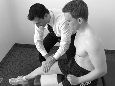
Superior Lateral Patella Adjustment: (See Photo 2)
- Patient: Supine, affected knee placed in the cervical headpiece, or on toggle board.
- Doctor: On affected side, facing caudad.
- Table: Cervical piece in the ready position, or toggle board under affected knee.
- Contact: Web contact on superior aspect of patella.
- Stabilize: Contact hand.
- LOC: S-I and L-M.
- The doctor must note that the superior lateral patella subluxation is often associated with a condition called Excessive Lateral Pressure Syndrome.
- This syndrome causes knee pain due to lateral tracking of the patella.
- When the patella tracts laterally, it results in the lateral facet of the patella grinding excessively against the lateral femoral condyle.
- This results in the destruction of the hyaline cartilage and roughening of the joint space.
 |
|
| Photo 2: Contacts for a superior lateral patella subluxation are displayed on a patient. Notice how the cervical headpiece is used as the dropping mechanism.
|
Following correction of the ilium and patellar subluxations, an exercise program for the vastus medialis should be implemented to eliminate future recurrence of this problem. The doctor can refer this to a personal trainer or can perform the rehabilitation within the clinic if he/she so chooses.
An interesting study from Montreal explored the vastus medialis muscle in great detail through dissection and electromyography.1 Researchers found that three separate groups of fibres existed within the vastus medialis: proximal fibres, medial fibres and distal fibres. The study stated that the proximal and medial fibres were inserted on a tendon common to the rectus femoris, whereas the distal fibres were attached directly to the medial aspect of the patella. Furthermore, the proximal fibres were significantly more active than the distal fibres during maximum knee extensions.1 The study concludes by stating that the role of the proximal fibres is one of assisting the rectus femoris in knee extension, whereas the sole purpose of the distal fibres is to track the patella medially.1 Therefore, when patients present with a superior lateral patella due to a weak vastus medialis, the rehabilitation program for the vastus medialis should focus on strengthening the distal fibres.
As usual, I have only scratched the surface on knee injuries and subluxations. The knee is complicated and problems can originate from the spine, the knee complex itself or the supporting musculature that affects it. The above case is only one potential problem that must be assessed if a knee issue exists. If you have any questions or concerns, please contact me at johnminardi@hotmail.com.
Until next time . . . adjust with confidence!
Reference
1. Lefebvre, R et al. Vastus medialis: anatomical and funtional considerations and implications based upon human and cadaveric studies. JMPT 2006; 29(2):139-144.
Dr. John Minardi is a 2001 graduate of Canadian Memorial Chiropractic
College. A Thompson-certified practitioner and instructor, he is the
creator of the Thompson Technique Seminar Series and author of The
Complete Thompson Textbook – Minardi Integrated Systems. In addition to
his busy lecture schedule, Dr. Minardi operates a successful private
practice in Oakville, Ontario. E-mail johnminardi@hotmail.com, or visit www.ThompsonChiropracticTechnique.com .
Print this page