
A 40-year-old female office worker presents with jaw pain. The patient
notifies the doctor that she underwent a root canal last week, during
which her mouth remained open for several hours while the dentist
performed the procedure.
CASE STUDY
A 40-year-old female office worker presents with jaw pain. The patient notifies the doctor that she underwent a root canal last week, during which her mouth remained open for several hours while the dentist performed the procedure. The patient states that since the procedure, she has been experiencing pain and clicking bilaterally at the temporomandibular joints (TMJ), as well as occasional tinnitus. The pain is aggravated during chewing, and relieved with intermittent application of ice to the affected area. Physical examination reveals pain and tenderness palpated at the TMJ and surrounding musculature bilaterally. The doctor also clinically observes that the patient is unable to place three fingers in their mouth. Furthermore, the doctor observes the patient’s mandible deviating to the right upon opening of the mouth. Neurological, and X-ray analyses are unremarkable.
 |
|
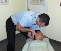
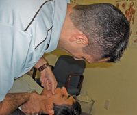
| Picture 1: Manual adjustment contacts for a right mandibular deviation, Part 1. Deviation side up, thumb pad contact on mandibular ramus, thrusting S-I with the slope of the articular eminence. Advertisement
|
This type of clinical presentation is the typical scenario that indicates suffering due to a mandibular subluxation through the TMJ. In this edition of Technique Toolbox, I will discuss the biomechanics of the TMJ, and how to correct for both the osseous component, as well as the muscular component involved in this injury. Rather than focus on a specific technique protocol, I will discuss a couple of options that a chiropractor can use to correct for the same biomechanical lesions, regardless of the method they choose to use.
BIOMECHANICS OF THE TMJ
The doctor must be aware that during opening of the mouth, normal biomechanics through the TMJ occur as follows:
- The first motion to occur is rotation, as the head of the mandible rotates on the meniscoid tissue.
- The second motion to occur is translation, as the meniscoid tissue translates on the articular eminence.
A subluxation involving the TMJ indicates that a problem exists with both sides of the jaw. However, the subluxation patterns are different from one side to the other.
- For example, if a patient exhibits right deviation with jaw opening, this indicates that the right TMJ is able to rotate, but cannot translate. Furthermore, the left TMJ is able to translate; however, it cannot rotate.
- In this example, the side of deviation (right side) is considered hypomobile, indicating that the subluxated mandible is jammed within the joint, with very little muscle involvement.
- However, the opposite side of deviation (left side) is considered hypermobile, indicating that a large muscular component is involved with the translational subluxation of the mandible.
|
|
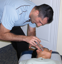
| Picture 2: Alternative Adjustment, using the Activator Instrument, for Part 1. |
STEP ONE – MANDIBULAR CORRECTION: TWO-PART ADJUSTMENT
Part 1: (See pictures 1 and 2)
Patient – Supine. Deviation side up.
Doctor – Head of table.
Contact – Thumb pad or Activator Instrument on mandibular ramus.
Stabilize – Cradling head, or stabilizing mandible.
Line of Correction (LOC) – S-I, in line with ramus of the mandible, or with slope of articular eminence.
The first part of this adjustment decreases the compression involved in the TMJ, and allows translation to resume.
|
|
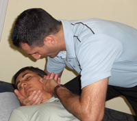
| Picture 3: Manual adjustment contact, Part 2. Non-deviation side up, thumb pad contact on mandibular condyle, thrusting I-S with the slope of the articular eminence. |
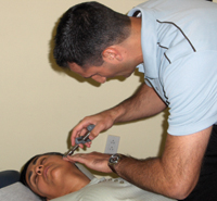
Part 2: (See pictures 3 and 4)
Patient – Supine. Deviation side down.
Doctor – Head of table.
Contact – Thumb pad or Activator Instrument on mandibular condyle.
Stabilize – Mandible.
LOC – I-S, in line with ramus of mandible, or with slope of articular eminence.
The second part of this adjustment will reduce the translation and allow rotation to resume.
STEP TWO – MUSCULAR COMPONENT
(See picture 5)
There are several muscles involved in the opening and closing of the mouth. Masseter, lateral pterygoid, medial pterygoid, and temporalis all play a role. Although it is very difficult to pin point which exact muscle will provide the most benefit, soft tissue therapy of these muscles will certainly provide a therapeutic benefit for TMJ disorders. It was originally thought that the lateral pterygoid could be accurately palpated and treated. However, other trains of thought have suggested that this muscle may not be easily palpated, and thus medial pterygoid and temporalis may indeed be the contact points.
 |
|
| Picture 4: Alternative Adjustment, using the Activator Instrument, for Part 2.
|
Therefore, on the hypermobile side (opposite side of deviation), the doctor will perform myofacial release on the medial pterygoid or temporalis.
Patient – Supine. Head in neutral position.
Doctor – Head of table.
Contact – Finger pad on medial pterygoid or temporalis inside the mouth.
Stabilize – Thumb pad stabilizes muscle from outside the mouth.
LOC – Sustained pressure contact until tension has released.
The doctor may also perform trigger point therapy, or Active Release Technique (ART), on the affected musculature – any of these would be effective.
Following the adjustment and muscular treatment, the patient should be able to place three fingers into the mouth, and the deviation should be significantly reduced.
 |
|
| Picture 5: Index finger pad contact on medial pterygoid or temporalis inside the mouth, on the hypermobile (non deviation) side.
|
Similar to all extremities, the TMJ must be assessed and corrected after the spine has been checked and all necessary subluxations have been adjusted. Ancantara et al. discusses a patient with bilateral ear pain, tinnitus, vertigo and headaches. The patient’s complaints were attributed to a diagnosis of TMJ syndrome, and were treated unsuccessfully. An atlas sub luxation was detected and corrected in the patient, which resolved the persistent symptoms in nine visits.1 The primary subluxation in this case was the atlas, which assimilated TMJ symptoms. This case reinforces the principle that the spine must be assessed and corrected prior to extremity adjusting. Other research has found that the majority of individuals who suffer from a TMJ disorder will also suffer from at least one otologic complaint.2 Otalgia, tinnitus, vertigo, and hearing loss were reported most frequently in patients suffering with TMJ disorder, whereas individuals lacking TMJ disorder had a much lower incidence of these symptoms.2 Therefore, if a patient presents with otologic symptoms, the doctor must be sure to rule out any potential problems with the TMJ, and correct them as necessary.
By using a combination of adjustments with muscle procedures, as in this case study, TMJ injuries can be treated efficiently and effectively. As demonstrated, provided that proper biomechanics are followed, manual adjusting, an activator instrument, or any other adjusting device can be utilized. Similarly, any effective soft tissue technique can be utilized to treat the muscular component. As usual, I have only scratched the surface with TMJ injuries. There are several other problems that can present, in which alternative treatments must be implemented. Don’t be intimidated by TMJ complaints – instead, educate yourself and be confident.
If you would like to see a specific technique featured in a future edition of Technique Toolbox, please e-mail me at johnminardi@hotmail.com.
Until next time . . . Adjust with Confidence! •
References:
- Ancantara, J. et al. Chiropractic care for patients with temporomandibular disorders and the atlas subluxation. JMPT. 2002. 25(1):63-70.
- Tuz, H. et al. Prevalence of otologic complaints in patients with temporomandibular disorders. Am J Orthod Dentofacial Orthop. 2003. 123(6):620-623.
Dr. John Minardi is a 2001 graduate of Canadian Memorial Chiropractic College. A Thompson certified practitioner and instructor, he is the creator of the Thompson Technique Seminar Series and author of The Complete Thompson Textbook – Minardi Integrated Systems. In addition to his busy lecture schedule, Dr. Minardi operates a successful private practice in Oakville, Ontario. E-mail johnminardi@hotmail.com, or visit www.ThompsonChiropracticTechnique.com .
Print this page