 My sincere thanks to my dear friend, colleague and classmate, Dr. Howard Fisher of Toronto, for providing me with this case.
This 43-year-old woman presented with dull, achy sub-occipital pain, with decreased upper cervical ranges of motion.
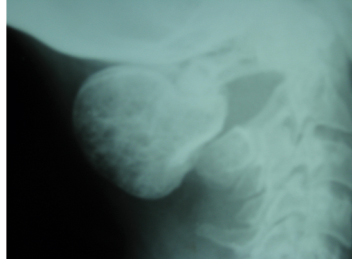
The radiograph reveals a markedly enlarged posterior tubercle of atlas (5 cm in diameter), with abnormal internal trabeculation.
Diagnosis: Osteoblastoma
Discussion:
– rare and benign; accounts for one per cent of all primary bone tumours
– originally described as an “osteogenic fibroma of bone” in 1956; shares similar clinical and histological features with osteoid osteoma, giant cell tumour and fibrous dysplasia
– commonly affects the vertebral column; 30 per cent in the posterior elements of the spine; also found in tubular bones
– bone-forming lesion (many immature bony trabeculae, lined with osteoblasts, demonstrating various degrees of ossification)
– may be cortical or medullary; when cortical, expansion is often present; may reach up to 11 cm in diameter!! (average 3.2 cm)
– may present with neurological symptoms as a result of cord or nerve root compression
– no helpful lab tests; biopsy required to confirm diagnosis
On X-ray:
– usually well-circumscribed cortical lesion, with thin shell of peripheral new bone
– lesion larger than 2 cm
– internally, displays varying degrees of radiolucency depending on amount of ossification of the numerous immature trabeculae
– may sometimes appear malignant, with adjacent cortical destruction and extraosseous soft tissue expansion
– CT is helpful in the management of the lesion, to provide accurate information regarding the size and extent of the lesion
Therapy:
– surgical resection; this is usually curative with good outcome
A 1983 CMCC graduate, Dr. Marshall Deltoff, DC, DACBR, FCCR(C), completed his radiology residency at Los Angeles College of Chiropractic. He is a past radiology department
chairman and residency coordinator at CMCC, and he initiated the
radiology curriculum at UQTR. Dr. Deltoff has lectured throughout North
America, and is co-author, along with Dr. Peter Kogon, DACBR, of the
radiology text “The Portable Skeletal X-ray Library” published by
Mosby-Yearbook of St. Louis. Dr. Deltoff can be reached at:
Images Radiology Consultants,
16 York Mills Road, Toronto,
ON M2P 2E5
Tel: (416) 512-2225
Fax: (416) 512-2226
e-mail: marshdel@rogers.com
Print this page