In this issue, we will review the main classification of diaphragmatic hernias:
In this issue, we will review the main classification of diaphragmatic hernias:
1. Esophageal hernia
• herniation of all or part of the stomach through the esophageal hiatus into the thorax
• the most common diagnostic entity identified on X-ray of the upper gastrointestinal tract
• found to some degree in 10 per cent of the adult North American population
• “sliding” type, involving the esophogastric junction and a portion of the gastric fundus; accounts for 95 per cent of these hernias
• “paraesophageal” type, in which the junction remains fixed below the diaphragm, with only a portion of the fundus herniating; represents only 1 per cent of hiatal hernias
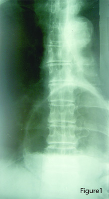
• hernia produces a mass shadow at the left medial base
• often can visualize air/fluid level in the intrathoracic portion of the stomach superimposed over the cardiac silhouette
• this permits diagnosis on plain film (Figure 1)

 • if not, the abnormality can be readily identified with a barium swallow
• the barium swallow also helps to differentiate this lesion from a pulmonary cyst or abscess, or a diaphragmatic tumour
• associated symptoms of reflux are usually the only clinical significance
2. Foramen of Morgagni hernia
• rare
• basal mass shadow
• usually small, containing some omentum and portion of bowel
• barium enema study may show upward angulation of the midtransverse colon
• pyloric region of stomach and proximal duodenum may be displaced upward
• in infants and young children, the liver may rarely herniate through the foramen of Morgagni
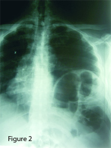
3. Foramen of Bochdalek hernia (Figure 2)
• herniation through the pleuroperitoneal foramen of Bochdalek
• often large herniation
• loops of bowel are visualized
• occasionally accompanied by left kidney
4. Traumatic herniation
• from severe crushing types of injury to the thorax or abdomen
• diaphragm may be ruptured completely, with defects in the parietal pleura and peritoneum
• left diaphragm involved in 95 per cent of cases
• if necessary, barium meal or enema may aid in identifying the portions of the gastrointestinal tract within the hernia
• MRI examination will also help clarify gastrointestinal status
References:
1. Juhl JH, Paul LW. The essentials of Roentgen interpretation, 3rd edition. Hagerstown, Maryland: Harper & Row, 1972.
2. Harger B, Hoffman L., in Clinical imaging with skeletal, chest and abdomen pattern differentials, Marchiori D and McLean I, eds, Chap 25-28, Mosby-Year Book, 1998.
Print this page