I would like to thank Dr. Robert Pollock, of Tottenham, Ontario, for sharing this case.
I would like to thank Dr. Robert Pollock, of Tottenham, Ontario, for sharing this case.
DIAGNOSIS:
os odontoideum
 |
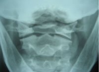
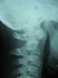
| Figures A (above) and B (below) clearly delineate a large superior ossicle representing the cephalad tip of the dens; there is absence of ossification of the body of the dens. |
 |
DISCUSSION:
– although odontoid anomalies are considered uncommon, this is the most frequently occurring anomaly of the odontoid process
– the apex and base of the dens fuse at approximately nine years of age, but the base does not fuse to the body of C2 until adulthood; this is normal
– the cephalic portion of the dens develops normally from its two lateral ossification centres, but remains ununited with the body of C2 above the level of the growth plate (the neurocentral synchondrosis)
– since the osseous defect is not found at the site of the growth plate, it has been hypothesized that os odontoideum is actually a longstanding and unrecognized fracture of the base of the odontoid; however, many authors still consider this entity to be a congenital nonunion
– may be associated with Down syndrome, Klippel Feil syndrome
RADIOLOGICAL FINDINGS:
– characterized by a round ossicle that may vary in size
– a smooth, sclerotic border separates the os from the axis
– the gap is wide with smooth margins; this distinguishes it from an acute odontoid fracture, in which the gap is typically narrow with irregular or jagged margins
 |
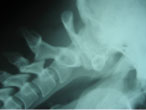
| Figure C demonstrates anterior translation of atlas on axis, with no change in the atlantodental interspace, signifying an intact transverse ligament in this woman. |
– another differentiating clue is an angular deformity of the posterior aspect of the atlas anterior tubercle (see Figures B and C)
– anterior atlas tubercle usually demonstrates stress hypertrophy
– odontoid tip may tilt on flexion/extension (see Figure C)
– transverse ligament is usually intact; no change in ADI on flexion
– posterior arch of atlas may be hypoplastic or absent as well
CLINICAL FINDINGS:
– patient is usually asymptomatic
– instability and complications resulting from relatively insignificant trauma pose a serious health risk
– paralysis or even death may result from trivial trauma or even spinal manipulative therapy
– prophylactic stabilization in the asymptomatic patient is still controversial, with surgical ntervention in cases of instability or in the presence of severe neurological manifestations
References:
Deltoff MN, Kogon PL. The Portable Skeletal X-ray Library. 1998, Mosby-Yearbook, St. Louis.Yochum TR, Rowe LJ. Essentials of Skeletal Radiology. 1987, Williams & Wilkins, Baltimore.
A 1983 CMCC graduate, Marshall Deltoff, DC, DACBR, FCCR(C) completed his radiology residency at Los Angeles College of Chiropractic. He is a past radiology department chairman and residency coordinator at CMCC, and he initiated the radiology curriculum at UQTR. Dr. Deltoff has lectured throughout North America, and is co-author, along with Dr. Peter Kogon, DACBR, of the radiology text “The Portable Skeletal X-ray Library” published by Mosby-Yearbook of St. Louis. Dr. Deltoff can be reached at:
Images Radiology Consultants,
16 York Mills Road,
Toronto, Ont. M2P 2E5
Tel: (416) 512-2225
Fax: (416) 512-2226
e-mail: marshdel@rogers.com
Print this page